Laboratory diagnostics &
cell science
Since 1968
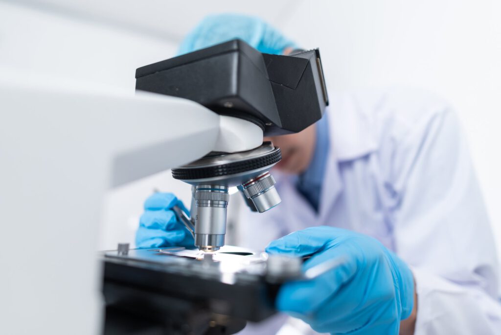
My fields of work and interest cover laboratory diagnostics and cell research. At this, immunology, immunohematology, blood transfusion, as well as quality management in medical departments are most important for me. My activities cover concepts to optimize and standardize processes to improve operating procedures, performances and services (e.g. laboratory analytics) on the basis of standards for the purpose of extended customer satisfaction.
Apart from laboratory diagnostics and respective activities in QM systems, my other fields of expertise include microscopy, immunohistology and cell research.
I would be happy to get in touch – please do not hesitate to send technical inquiries, information, and suggestions my way:
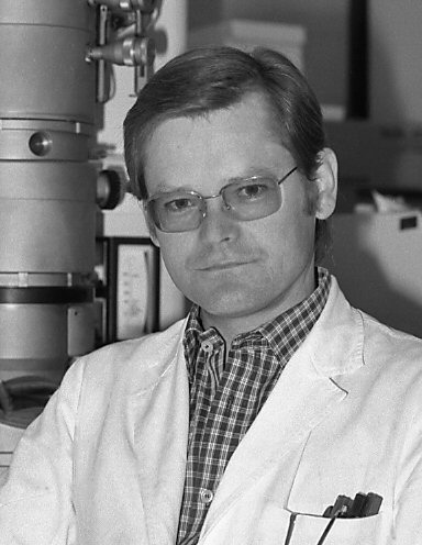
Prof. Dr. med. Wolf D. Kuhlmann
Laboratory Medicine & Cell Science
kuhlmann@kuhlmann-biomed.de
Education & grants for research
Education
Elementary and high school in Siegen, Germany
Study of Medicine, Faculty of Medicine University of Heidelberg
Doctorate M.D, Faculty of Medicine University of Heidelberg
Doctoral thesis: Immunochemical and immunohistological studies of normal and atherosclerotic altered blood vessels in humans (summa cum laude)
Postdoctoral qualification for a teaching career in higher education at the Faculty of Medicine, University of Heidelberg, Habilitation and College lecturer (venia legendi)
Habilitation treatise: Electron microscopic immunocytochemistry: Peroxidase as marker and labeling reagent for immuno-enzyme detection of antigens and antibodies
Other certifications: Licence to practice medicine; specialist in laboratory medicine, blood Transfusion
Medical council: Bezirksärztekammer Koblenz
Landesärztekammer Rheinland-Pfalz
Grants for research
World Health Organization (WHO)
Deutsche Forschungsgemeinschaft (DFG)
Heisenberg-fellowship (DFG)
Sonderforschungsbereich Krebsforschung (DFG, SBF136)
Ministry of Defense (BMVg)
Scientific awards & historical notations
Historical notations
Scientific advice in histochemistry started from my biomedical projects at the DKFZ (Deutsches Krebsforschungszentrum in Heidelberg).
Work on antibody labeling with enzymes encouraged the idea to make available immunohistochemical instructions and special reagents for scientists. The intention in doing such things, however, goes back to the time when I was studying medicine and being busy in immunofluorescent cell labeling for my doctoral Thesis.
Scientific awards
Robert-Schwank-Preis (1969)
Robert-Feulgen-Preis (1976)
Professional career &
main places of work
Medical Clinic (Ludolf-Krehl-Klinik) University of Heidelberg
Institut de Recherches Scientifiques sur le Cancer (IRSC/CNRS) Villejuif
German Cancer Research Center (DKFZ) Heidelberg
Ernst-Rodenwaldt-Institut Koblenz, Central Institute of the Armed Medical Forces, Ministry of Defense
Labor Koblenz MVZ für Laboratoriumsmedizin Koblenz-Mittelrhein
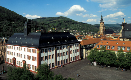
University of Heidelberg
Doctoral Thesis and how my career began at the Ludolf-Krehl-Klinik in Heidelberg
Interest in research and basic laboratory techniques came into shape in 1964 when I started my doctoral thesis in the LUDOLF-KREHL-KLINIK (Medizinische Universitätsklinik Heidelberg, chair Prof. Dr. G. Schettler) on the subject: Immunochemical and immunohistological studies of normal and atherosclerotic altered blood vessels in humans. At that time, the medical clinic established the research unit Immunobiology with projects in the analysis of human stomach mucosa. The starting team was small (Dr. W. RAPP, one technician and I myself) and inspired by the early works from the Institute of Experimental Cancer Research in Heidelberg (established 1906 as independent department at the CZERNY-KLINIK and founded by V. CZERNY for cancer patients). The very beginning of immunology in Heidelberg can be traced back to the scientists E. VON DUNGERN, L. HIRSCHFELD (L. Hirszfeld), H. SACHS and E. WITEBSKY with their works on serologic specificity of organs (normal, tumor and embryonic tissues). Also, stimuli are given by works of P. GRABAR and staff members at the Institut de Recherches Scientifiques sur le Cancer in Villejuif on the immunoelectrophoretic analysis of biomolecules.
During the first months we housed in a patient’s room of the Medical University Clinic (Station Naunyn). Technical equipment was sponsored by the DFG (Deutsche Forschungsgemeinschaft) and the LUDOLF-KREHL-KLINIK. Within few months the available space became insufficient. Thus, we moved into a garden shed of of the clinic park for about two years. Our research focus was on new techniques and know-how in immunology, biochemistry and histology. During that time, a new laboratory building for clinical research was built up with enough workspace for all research groups of the hospital. The LUDOLF-KREHL-KLINIK provided equipment for immunofluorescence as an important tool in diagnostics of autoimmune diseases. This helped our research work gain traction. Moreover, we could invest in the acquisition of an electron microscope.
In these days, laboratory work in purification of biomolecules, preparation of antibodies by immunization and biochemical characterization was a feat of skill. Time-consuming steps were necessary to obtain enough purified antigens, specific antibodies and marker labeled antibodies for application either in analytics or in cell science studies. Hence, the experimental efforts required much time over several years including works at many nights and weekends. I still remember that literature research, literature supply and purchase of fine chemicals were extremely tedious because computer-based search on the Internet did not exist. Besides, photocopiers were not omnipresent and on top, personal computers with text processing software were not available at that time; all written work done with a conventional typewriter.
Research and studies on immunohistology and cell Research by electron microcopy at the Institut de Recherches Scientifiques sur le Cancer in Villejuif
During my doctoral thesis I stayed at the Institut de Recherches Scientifiques sur le Cancer in Villejuif (France) several times to work on immunochemical, immunofluorescence and enzyme-cytochemical methods. I valued the spirit, the open-minded atmosphere of this research institute and the meetings with prominent persons like P. Grabar, P. Burtin and A. Lwoff. When finishing my thesis, I moved for some years to the Institut de Recherches Scientifiques sur le Cancer in Villejuif for research on immuno-enzyme techniques and selective cell labelings at the electron microscopic level. It was a great opportunity for me being in the laboratories of Dr. W. BERNHARD, Dr. E. H. LEDUC, Dr. S. AVRAMEAS and Dr. J. URIEL, the worthy pioneers in electron microscopy and immunochemistry, respectively. Dr. W. BERNHARD deemed as Nestor of cell biology not only in France but also in all of Europe.
In Villejuif originated at that time many papers on ultrastructural cytochemistry and immunohistology. I appreciated very much the helpful advice of A. VIRON in electron microscopic techniques and in particular the cooperation in the development of techniques for cryo-ultramicrotomy. Then, antigen and antibody labeling techniques with marker enzymes such as horseradish peroxidase and glucose oxidase from Aspergillus niger were special issues. In collaboration with M. BOUTEILLE, E. H. LEDUC, S. AVRAMEAS and H. R. P. MILLER, we published our findings on differentiation und maturation of lymphocytes following antigen stimulation (enzymes as antigens and markers). Combined electron microscopy, immuno-staining and autoradiography with 3H-thymidine proved well suited to reveal ultrastructure and antibody formation in the course of cell proliferation, differentiation and maturation during the immune response.
It was interesting to observe that immuno-enzyme techniques developed rapidly in Europe, while ferritin labeling was still in fashion in USA and other countries. This became obvious at the Gordon Research Conference in 1972 (Immuno-Electron Microscopy, Wayland Academy, Beaver Dam). In any case, immuno-enzyme techniques have become most valuable in biomedicine. It is worth mentioning that the principles of immuno-enzyme techniques are still widely applied tools in cell biology. They also serve as a platform for the detection of biomarkers in a large number of diagnostic settings and in the detection of molecular probes in many Sciences.
Scientific Projects on alpha-Fetoprotein, hepatocarcinogenesis and cell differentiation at the Deutsches Krebsforschungszentrum in Heidelberg
Coming back to Heidelberg, I was engaged for many years with intensive research at the Deutsches Krebsforschungszentrum Heidelberg (DKFZ, Institut für Nuklearmedizin). Projects covered studies on alpha-1-fetoprotein as bio-marker for liver regeneration following different forms of injuries and for the expression of an oncodevelopmental gene product during hepatocarcinogenesis; all by grants of the Deutsche Forschungsgemeinschaft (DFG, Sonderforschungsbereich 136 Krebsforschung). At work, cooperation came across with Prof. W. KUNZ from the Department of Biochemistry (Regulation and expression of microsomal enzymes in the course of chemical hepatocarcinogenesis). Furthermore, studies were done with Prof. K. H. WURSTER from the Institute of Pathology and with Prof. W. RAPP from the LUDOLF-KREHL-KLINIK on Gastrointestinal glycoproteins as marker in histogenesis and histopathology. All projects yielded new biochemical and histochemical methods. In parallel, I assisted the laboratory of Immunobiology of the LUDOLF-KREHL-KLINIK in diagnostic issues: proof of paraproteins (immunofixation, immunoelectrophoresis), measurement of serum proteins (nephelometry, immunodiffusion according to Mancini and Oudin), detection of autoantibodies in patients suspect of collagenosis (ANA immunofluorescence, ANA/ENA differentiation by Ouchterlony’s gel diffusion techniques and by counter current immunoelectrophoresis).
The mission of finding the hepatic progenitor cells and AFP expression in liver injury and hepatocellular carcinoma has taken many efforts. The expression of this oncodevelopmental gene product in ontogeny and liver regeneration proved to be suitable as marker as marker for questions as to the role of putative progenitor and stem cells in liver disease. The experimental models included partial hepatectomy, acute injury by carbontetrachloride, D-galactosamine (GalN) and N-nitrosomorpholine (NNM) as well as protocols of chemical hepatocarcinogenesis by feeding of NNM in low doses. Serological and immunohistological detection of AFP gene expression served to follow pathways of cellular differentiation. To this aim, I had to acquire considerable skills, especially for the light and electron microscopic studies with tissue preparation such as fixation, embedding, cryo-techniques and staining with selective reagents. Major challenges concerned immuno-staining of specimens for electron microscopy.
Many experiments let me conclude that under certain conditions, in which the proliferative capacity of normal adult hepatocytes is blocked, stem-like cell populations replaced loss of liver mass. The progeny of the stem cell compartment (bipotential progenitor cells) proliferated and differentiated into hepatocytes and bile duct epithelia. Comparable to other organ systems, lineage cells may consist of short-term and long-term stem cells, precursor cells, and mature cells.
Interestingly, no stem cells were required in models of surgical removal of parenchyma or in carbon tetrachloride intoxication while regeneration of injured liver by GalN and NNM occurred through proliferation of biliary epithelial cells. These biliary epithelial cells, collectively called oval cells, are derived from the canals of HERING. Proliferating bile duct cells reached levels of differentiation with reactivation of fetal genes and AFP synthesis rated as sign of certain degree of retrodifferentiation and potential stemness. Due to the same embryonic origin of bile ducts and hepatocytes, biliary epithelium and its proliferating progeny (oval cells) must have a defined role in liver regeneration as a transit and amplification compartment. Certainly, factors of the liver microenvironment have also an impact on the fate of progenitor cells with respect to differentiation into either hepatocytes or bile ducts.
Malignancy may arise from proliferating/differentiating oval cells or from adult hepatocytes; both cell types have stem-like properties. Oval cells will function as facultative progenitors for hepatocytes and biliary tract cells. In the experimental design, they can also give rise to regenerated hepatocytes with high risk for transformation and being foci of altered hepatocytes. Finally, AFP-positive and AFP-negative carcinomas occurred in the same liver. This may represent random clonal origin through genetic and epigenetic alterations and AFP resurgence as a process of retrodifferentiation.
Scaling up laboratory diagnotics and technical developments of the Medical Corps of the German Armed Forces, Ernst-Rodenwaldt-Institut in Koblenz
At the end of the 1980’s, my fields of activity changed. There were new projects in laboratory medicine in my new chosen environment at the ERNST-RODENWALDT-INSTITUT (Hygienisch-Medizinisches Institut, Zentrales Institut des Sanitätsdienstes der Bundeswehr Koblenz). This military institution with tradition in hygiene, infectious diseases and health care is named after the renowned hygienist and specialist in tropical diseases E. RODENWALDT (emeritus professor of the Medical Hygiene Institute, University of Heidelberg, and father of geomedicine). In providing laboratory diagnostics at a comprehensive scale, my initiatives focused on techniques such as nephelometry, ELISA, TR-FIA, RIA, MLCT and PCR for HLA-typing, granulocyte function tests (oxidative burst), flow cytometry (lymphocyte subset typing) and quantitative assays for antibodies related to vaccination. The latter served also for sero-epidemiological studies. PCR and DNA/RNA hybridization techniques were also applied as useful tools in laboratory diagnostics.
Handling of patient probes and laboratory workflow were still old-fashioned and error-prone at that time. With the availability of computers things changed gradually. We first experimented with MS-DOS (x86-based PC) and programming in Microsoft BASIC to improve laboratory workflow. However, real improvement first became possible with the subsequently introduced UNIX based laboratory information and management system (LIMS, HP-UX). Then, the knowledge-based expert system Pro.M.D. shell (Chr. Trendelenburg and B. Pohl) was adapted for evaluation and reporting of test results. This system based on PROLOG-2 (Expert-Systems Ltd., Oxford, England) was crucially adapted by coworker Dr. J. P. SCHRÖDER. It proved useful in the interpretation of antibody titers giving tips for booster vaccinations or warnings of allergic reactions. By the time, another concept called LIPS framework (Learning, Information, and Performance Support) came in use for such work. We realized some projects on vaccine immunization and sero-epidemiologic features which were published together with Dr. J. P. SCHRÖDER and DR. J. RIEGER. Several meetings of the Arbeitskreis Immunprophylaxe organized by Dr. M. PIETSCH were of great interest because of exchange of expertise with other scientists working in the field of vaccination. Military vaccination schedules provided us with thousands of serum probes to quantify serum antibodies. The question of possible type I and type III immune reactions in vaccinated persons let us have access to respective case histories. Indeed, we found in some rare cases IgE mediated sensitization by tetanus toxoid vaccine. Serum specimens of highly positive cases enabled us to develop an in vitro test system for suspected IgE sensitization (allergic reaction type I) following tetanus toxoid booster immunizations.
I want to mention the very valuable cooperation with Prof. A. MARKEWITZ and Prof. A. FRANKE (Bundeswehr Zentralkrankenhaus) and the participation in their research projects Humoral and cellular immunity following cardiac operation. The papers on humoral and cellular immunity, on inflammatory and anti-inflammatory cytokine networking in patients after surgical interventions were of high quality, reflected by high citations rates.
In the wake of the Iron Curtain’s fall in Central Europe much shedding of military facilities was planned and indeed realized. Interestingly, military administration’s first proposal was to establish four new institutes (sic!) by reorganization of the one existing Central Institute in Koblenz. This project, however, was a venture without reference to reality because four institutes would be a too ambitious project for a shrinking army. At the instigation of politics, the German Army suffered soon from cannibalization effects. Many pieces of filet were cut away while only residuals were left from the tried-and-true Central Institute in Koblenz. The Department of Immunology should remain as a laboratory group with three laboratory assistants and one physician. In doing so, the Department of Immunology and its attractive facilities according to STAN (the so-called strength and equipment standard) with four laboratory units, a total of 5 medical doctors and 13 medical-laboratory assistants was melted down. The many changing decisions in restructuring the Central Institute were exhaustive and an example for the never-ending political quarrels in the German federal republic at the time. Lacking foresight, decision-makers did not have on their radar the general importance of immunology in medicine and life science. Staff was damned to resign, team members transferred from here to there and some to the military hospital in Koblenz.
Laboratory medicine in the environment of a business-driven medical practice, Labor Koblenz
Today, I work with former medical officers, who have founded their own medical center for laboratory medicine (MVZ für Laboratoriumsmedizin Koblenz-Mittelrhein, Labor Koblenz). Labor Koblenz is quite different from the institutions so far described. Though laboratory analyses are the main subject, the MVZ turned out to be a laboratory with a significantly larger analytic spectrum and with significantly higher numbers of analyses per day as compared with any other lab, in which I worked before. Features that shape the business decisively are the confined hierarchies, then items and rules of statutory and private health insurances as well as the idiosyncrasies of discounted price setting for the acquisition of new orders from hospitals (all risks whatsoever). Diligent calculations of costs and profit are called for. One can believe that some modern organizational structure according to industrial models will be more effective than retaining the conservative charm of a medical practice in order to settle properly matters such as personal structure, allocation of responsibilities, duties and benefits.
Large quantities of patient specimens are processed every day. It is unbelievable however, how much time sample reception is spent on inadequate or false notations which requires numerous phone calls to clarify with clients. Also new to me in this facet of laboratory medicine were the daily vexations due to sometimes poor sampling quality of patient probes at the primordial locality (medical practice) as well as the laxity of courier services in transport conditions. All these are critical aspects of high quality before the actual laboratory analytics even begin. Analytics themselves are usually correct due to strict quality controls and accreditation standards.
Within this setting, I mainly deal with autoimmunity, immunohematology and out-house activities. The latter includes care of external hospital laboratories by controlling their workflow in clinical chemistry, immunohematology and blood transfusion. To that end, it was time to phase in quality management systems and audits. There are also audits in hemotherapy concerning safety in blood transfusion (monitoring of transfusion quality on behalf of the medical association of Rheinland-Pfalz). The organization of point-of-care testing (POCT) was a special job and not easy at all. Fostering sensitivity for quality insurance, training in the appropriate use of technical devices and documentation to satisfy laws and regulations were a long process. Now, POCT units in the hospitals supervised by us are on a good way.
Upcoming challenges the field of laboratory medicine will have to face
Analytic technologies have become more and more efficient but without invention of completely new ones. Laboratories depend particularly on precision, reproducibility and least possible deviance. Then, further automation and digitization are the next step to enable a fast, convenient and precise workflow. In the coming years, we will face some challenges in automation systems, software and communication ways that will change procedures in this business. Robots will take over a large share of the processing of laboratory work.
We see already trends in miniaturization of analytic systems. They are already in use in some diagnostic areas. High-performance POCT devices will compete with centralized analytics (classical medical chemistry, serology and microbiology). Lateral flow tests, POCT technologies using e.g. centrifugal-microfluidic chips for automated analysis, new marker conjugates as detection molecules, then new sensor, transducer and spectroscopic developments (Surface-Enhanced Raman Scattering and others) with highly developed microelectronics are likely candidates for specific and sensitive POCT diagnostics. This and the analysis of multiple parameters (multiplexing) will probably cross molecular genetics.
Then, we will have to familiarize ourselves with computer algorithms and artificial intelligence (AI) that allow computer, machines and man to function in an intelligent manner. With immediate effect, bioinformatic approaches will change classification of diseases. Conventional protein-based biomarkers and new concepts like liquid biopsy/liquid profiling to characterize special disease features (ctDNA, RNA, miRNAs, SNPs etc.) are further stages in precision medicine.
Application of AI and neural networks in medicine are in rapid progress, e.g. in radiology, personalized diagnostics including histopathology and treatment of different diseases. All this is favorable, but it implies a considerable demand on physicians to acquire new skills and learn to include new and upcoming technology in their daily work. Without a doubt, we have to keep up with technological progress and with health requirements for our own good. Quite similar in meaning with the conception of CARL FRIEDRICH GAUSS:
„Wahrlich es ist nicht das Wissen, sondern das Lernen, nicht das Besitzen, sondern das Erwerben,
nicht das Da-Seyn [Dasein], sondern das Hinkommen, was den grössten Genuss gewährt“
(C. F. Gauss, XXX. Gauss – Bolyai. Göttingen, 1808, IX. 2. In: F. Schmidt, P. Stäckel (eds.), Briefwechsel zwischen Carl Friedrich Gauss und Wolfgang Bolyai, pp 93-94. Verlag B. G. Teubner, Leipzig 1899).