Laboratory diagnostics &
cell science
Since 1968
Laboratory medicine and cell research are typical areas of experimental medicine with the aim of understanding diseases and improving overall health.
Laboratory procedures are subject to constant improvement. They can prepare ways for precision medicine by tracking molecular and genetic characteristics in diseases. Quality-assured workflows are an important prerequisite for this goal.
Technical inquiries, informations and suggestions are welcome:
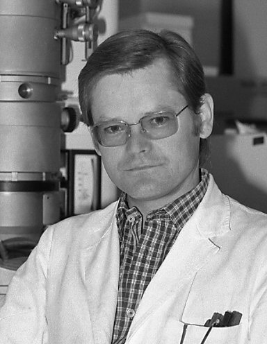
Prof. Dr. med. Wolf D. Kuhlmann
Laboratory Medicine & Cell Science
kuhlmann@kuhlmann-biomed.de
Education & Grants for Research
Education
Elementary and high school in Siegen, Germany
Study of medicine, Medical Faculty of Heidelberg University
Doctorate M.D., Medical Faculty of Heidelberg University
Doctoral thesis: Immunochemical and immunohistological studies of normal and atherosclerotic altered blood vessels in humans (summa cum laude)
Post-doc qualification for a scientific career: Habilitation in the field of experimental
medicine at the Medical Faculty of Heidelberg University and Venia Legendi
Habilitation treatise: Electron microscopic immunocytochemistry: Peroxidase as marker and labeling reagent for immuno-enzyme detection of antigens and antibodies
Other certifications: Licence to practice medicine; specialist in laboratory medicine, blood Transfusion
Medical council: Bezirksärztekammer Koblenz
Landesärztekammer Rheinland-Pfalz
Grants for research
World Health Organization (WHO)
Deutsche Forschungsgemeinschaft (DFG)
Heisenberg-fellowship (DFG)
Sonderforschungsbereich Krebsforschung (DFG, SBF136)
Ministry of Defense (BMVg)
Note in Advance
My interest in scientific subjects was already awakened and encouraged in my school days by our biology teacher Dr. Rombeck, who set up many working groups (biology), also in cooperation with the adult education center. Theoretical and practical aspects, applications in microscopy, histology and experimental biology were comprehensively covered with expert knowledge. This experience laid the foundation for studying medicine.
Many technical problems had to be solved during the experiments on labelling antibodies with fluorescent dyes for the localization of antigens in tissue sections. Thus, as well as in the later work in the field of immunochemistry and immunohistology, the plan matured to develop work instructions for special reagents and methods for cell biology issues. The result was “Histotechnik” and “Laboratory Diagnostics & Cell Science” (online and freely accessible).
Professional Career & Scientific Awards
Medical Clinic (Ludolf-Krehl-Klinik) University of Heidelberg
Institut de Recherches Scientifiques sur le Cancer (IRSC/CNRS) Villejuif
German Cancer Research Center (DKFZ) Heidelberg
Ernst-Rodenwaldt-Institut Koblenz, Central Institute of the Armed Medical Forces, Ministry of Defense
Labor Koblenz MVZ für Laboratoriumsmedizin Koblenz-Mittelrhein
Robert-Schwank-Preis (1969)
Robert-Feulgen-Preis (1976)
Heisenberg-Stipendium
Doctoral thesis & Professional Career, Ludolf-Krehl-Klinik, Heidelberg
When I started studying medicine, my interest in biomedical research increased. I was given the opportunity to work experimentally as a doctoral student at the Ludolf-Krehl-Klinik under the supervision of Prof. Dr. G. Schettler. The task was to carry out immunochemical and immunohistological studies on normal and atherosclerotic blood vessels in humans. However, neither suitable methods nor suitable premises were initially available for the experimental start. The clinic’s reference library was clinically oriented; literature research was laboriously carried out via interlibrary loan.
At that time, the Ludolf-Krehl-Klinik was pursuing the goal of setting up an immunobiology research group to work on projects involving the immunochemical analysis of human organs. The initial team consisted of three people: Dr. W. Rapp as the nominated head of the working group and recently returned from France from the Institut de Recherches Scientifiques sur le Cancer, then, a medical-technical assistant and myself a PhD student and newcomer. The working group “without space and equipment” started work in a patient room at the University Medical Center (Naunyn ward). The lack of space allowed a lot of time for the literature search. I drew my initial motivation from the early work of the Institute for Experimental Cancer Research in Heidelberg, which was founded as a department at the Czerny Klinik in 1906. The beginnings of immunology in Heidelberg, based on the work of E. von Dungern, L. Hirschfeld, H. Sachs and E. Witebsky were interesting, they already had the topic of serological organ specificity in mind. It was like an inspiration. After a few months, further impetus came from the working group led by P. Grabar and his colleagues at the Institut de Recherches Scientifiques sur le Cancer in Villejuif, but more on that later.
The research work was supported by both the German Research Foundation (DFG) and the Ludolf-Krehl-Klinik. The antiquated and spartan equipment improved continuously and facilitated our experiments. However, we soon reached the limits of the available space and were allowed to move into a solid garden house in the clinic park, which was available to us as a laboratory for about two years. During this time, the construction of a new laboratory building began, which was specially designed for clinical research at the Medical University Hospital.
The Ludolf-Krehl-Klinik supported us for many years and provided us with important equipment, including a fluorescence microscope for scientific and diagnostic purposes (autoimmune diseases). For histological examinations, we were allocated a microtome from the clinic’s old and stored stock (Hn 40 sledge microtome from R. Jung Nussloch) for the microtomy of paraffin-embedded tissue preparations. As we learned much later, it was a microtome from the time of Prof. L. Krehl, then head of the Krehl Clinic and subsequently head of pathology at the Kaiser Wilhelm Institute for Medical Research. Despite its age, this microtome was in excellent condition and is still in use for my histopathological work since decades. Over time, more and more machines and high-quality research equipment were added to the laboratory, and later an electron microscope with accessories. Since then, I have literally worked and lived in machine rooms.
The memories of the early days with all their hardships are still firmly anchored in my memory. The preparation of biomolecules, the production of antibodies by immunization and their biochemical characterization were time-consuming processes. The search for literature and the purchase of fine chemicals were laborious. At that time, there were no computerized Internet searches, photocopiers were not widely available and office computers with word processing software did not yet exist. All documents had to be written on conventional typewriters. It was a time that was characterized by manual work, perseverance and noisy machines.
Immunohistology, Cell research & Electron Microscopy, Institut de Recherches Scientifiques sur le Cancer, Villejuif
During my doctoral studies, I regularly visited the Institut de Recherches Scientifiques sur le Cancer in Villejuif (France) to study immunochemical and cytochemical methods. I greatly appreciated the atmosphere at this institute, as well as the encounters with personalities such as P. Grabar, P. Burtin and A. Lwoff. After completing my doctorate, I moved to this institute in Villejiuf for some years to work on immuno-enzyme techniques and selective cell labeling for electron microscopy. Finally, I considered it a privilege to be able to work in the laboratories of pioneers in the fields of immunochemistry, cell biology and electron microscopy (Dr. W. Bernhard, Dr. E. H. Leduc, Dr. S. Avrameas, Dr. J. Uriel). W. Bernhard in particular, the doyen of cell biology in France, was an inspiring personality.
In Villejuif, we contributed in many ways to electron microscopic cytochemistry and immunohistology. I greatly appreciated the collaboration with A. VIRON in the development of techniques for cryo-ultramicrotomy. At that time, various antigen and antibody labelling techniques were developed for the localisation of tissue structures using light and electron microscopy. The use of horseradish peroxidase took centre stage, especially as a suitable cytochemical detection method based on diaminobenzidine-H2O2 was available. This marked the beginning of a new period in immunohistochemistry, which can be described as the “peroxidase era”. Other enzymes such as alkaline phosphatase and glucose oxidase were quickly published as suitable markers, too. This enriched the field of immunoenzyme techniques. The use of marker enzymes was a new area of cell biology research. Numerous method developments began. Electron microscopy, immunoenzyme staining and autoradiography with ³H-thymidine enabled detailed visualisation of ultrastructure and cell function, for example the different developmental stages of lymphocytes during the immune response. Several experimental studies on the differentiation and maturation of immune cells after antigen stimulation were carried out in collaboration with colleagues M. Bouteille, E. H. Leduc, S. Avrameas and H. R. P. Miller. Immunoenzyme techniques have finally stimulated other areas of cell research and histopathology in a variety of ways.
It was striking how immunoenzyme techniques developed rapidly in Europe, while ferritin labeling was still predominant in the USA and other countries. This development became clear at the 1972 Gordon Research Conference (Immuno-Electron Microscopy, Wayland Academy, Beaver Dam). Over time, immunoenzyme techniques have developed into important tools in biomedicine. Today, they are the basis for the detection of marker molecules in diagnostic laboratories and for the detection of selective marker probes in molecular biology applications. The principles of immunoenzyme technology, its variants and diverse detection options are part of the toolkit of biological and diagnostic laboratories.
Cell differentiation, Biomarkers & Hepatocarcinogenesis, Deutsches Krebsforschungszentrum, Heidelberg
After returning to Heidelberg, we began projects at the DKFZ in Heidelberg (German Cancer Research Center Heidelberg) to investigate alpha-1-fetoprotein (AFP, an onco-fetal gene product) as a biomarker and indicator of the regenerative capacity of the liver after various types of damage. The expression of this onco-fetal gene product served as a marker for the cells involved and their differentiation stages in liver regeneration and during hepatocarcinogenesis. The appropriate methods for immunochemistry, serology, histopathology and immunohistology had yet to be developed.
The projects were funded by the German Research Foundation (DFG projects and Sonderforschungsbereich 136 Cancer Research). Joint work on the regulation and expression of microsomal enzymes in chemical hepatocarcinogenesis was carried out with Prof. W. Kunz from the Department of Biochemistry at the DKFZ. In addition, studies on the use of biochemically defined gastrointestinal glycoproteins as markers for histogenesis and histopathology were carried out with Prof. K. H. Wurster from the Institute of Pathology (University of Heidelberg) and Prof. W. Rapp from the Ludolf-Krehl-Klinik (Heidelberg). The production of special reagents for the detection and quantification of antigens/antibodies, but also the development of methods for light and electron microscopy were particular challenges. Ultimately, the effort involved in the search for precursor and stem cells was worthwhile.
From the many experiments it was deduced that under certain conditions the proliferative capacity of adult and differentiated hepatocytes is blocked and stem cell-like populations replace the loss of liver mass. The cells developing from the stem cell compartment (bipotent progenitor cells) are capable of differentiating into adult hepatocytes and bile duct epithelia.
After surgical removal of liver parenchyma, so-called stem cells are not required for regeneration. The remaining parenchyma is able to do this. This can also be seen after carbon tetrachloride poisoning. The situation is different after damage with GalN or NNM. Liver regeneration proceeds via the proliferation of biliary epithelia. These proliferating biliary epithelial cells, also known as “oval cells”, originate from the Hering ducts. They reach stages with reactivation of fetal genes and subsequent AFP synthesis, a process that is regarded as retrodifferentiation with potential stem cell properties. Bile ducts and hepatocytes have a common embryonic origin. Depending on the damage, the bile duct epithelium with its proliferating populations (oval cells) demonstrates its role as a transit and amplification compartment in the process of liver regeneration. It is assumed that additional factors in the hepatic microenvironment can influence the fate of the progenitor cells with regard to differentiation into liver cell epithelium (hepatocytes) and bile duct epithelium. Progenitor cells and adult hepatocytes have stem cell-like properties.
It is to be expected that genotoxic factors exert their effect both on proliferating oval cells with the populations differentiating from them and on the existing, differentiated adult liver cells. Observations suggest that oval cells can develop into regenerating liver cells with a high transformation risk as well as into focally altered liver cell areas (preneoplastic nodules). Both AFP-positive and AFP-negative carcinomas develop in one and the same liver. The phenomenon of AFP recurrence is generally regarded as a process of retrodifferentiation, possibly as a random clonal event due to genetic and epigenetic changes.
Parallel to the projects at the DKFZ, the habilitation procedure (experimental medicine) was prepared. At the same time, the immunobiology laboratory at the Ludolf-Krehl-Klinik was supervised: paraprotein diagnostics with immunofixation and immunoelectrophoresis, quantitative determination of defined serum proteins (nephelometry, immunodiffusion according to Mancini and Oudin), collagenosis diagnostics (detection, titer and microscopic differentiation of anti-nuclear autoantibodies) using indirect immunofluorescence. ENA differentiation was carried out using the usual methods at the time (agglutination and gel techniques).
Laboratory Diagnostics & Expansion of New Procedures, Central Medical Service of the German Army, Ernst-Rodenwaldt-Institut, Koblenz
After many years of research in experimental medicine, I expanded my field of activity. The new, attractive challenge was to add new procedures to the diagnostic repertoire in the Department of Immunology at the Ernst-Rodenwaldt-Institut in Koblenz (Central Institute of the German Armed Forces Medical Service). The military medical institute of the German Armed Forces was known nationwide for its long tradition in the fields of hygiene, infectious diseases and public health. Modern immunodiagnostics were the order of the day for military medical operations. There was also a focus on civil-military cooperation.
The institute revealed outdated structures in all work processes, from the delivery and recording of test samples to further processing (analysis, recording of measured values, documentation, preparation of findings, issuing of findings). The administrative processes (procurement, human resources, mail, etc.) were cumbersome. All in all, there was a high expenditure of personnel and time. The office technology was outdated and by no means up to date. The means of communication for cooperation with civilian agencies were simply an imposition.
Error-prone workflows in sample processing slowly changed with the availability of computers (PCs), which were supplied in small numbers from around 1990, suitable only for simple text processing. Real hardware and software support only came much later. In the early days, we ourselves started with MS-DOS (x86-based PCs) and programming in Microsoft BASIC to improve work processes. The conscripted soldiers were a great help (!). Progress came later with the introduction of a UNIX-based laboratory information and management system (LIMS, HP-UX). The courage to help ourselves was called for, because support for its use came only much later when material had been delivered several months before.
In the laboratories, the standard immunological methods (gel diffusion technique, agar immunoelectrophoresis, haemagglutination, turbidimetry) were gradually replaced by new techniques. Immunoassay systems were introduced, e.g. enzyme immunoassays (ELISA), fluorescence-based immunoassays (direct and indirect fluorochrome techniques, time-resolved TR-FIA fluorescence immunoassays) as a supplement to radioimmunoassays (RIA) for the measurement of hormones, drugs, tumor markers, autoantibodies, other serum/plasma components. In addition, cell-based assays have been established, including flow cytometry (FACS analysis) for leukocyte typing, cellular immunoassays for granulocyte function (phagocytosis, oxidative burst), serological typing of HLA-ABC and HLA-DR antigens (MLCT).
The introduction of molecular biological techniques (PCR-SSP/SSO, in-situ hybridisation, agarose gel electrophoresis) led to refined detection methods. Numerous new possibilities followed for research and diagnostic purposes. Molecular-specific tools offer a significant expansion of diagnostic possibilities. At the International Congress of Military Medecine in Rome (24.09.-26.09.1986), we presented the current state of development in the Department of Immunology at the Ernst-Rodenwaldt-Institut in Koblenz. The principles of ‘molecular probe mapping’ with examples of their application (hormones, proteins, virus components etc.) were demonstrated: Developments in immunology and the impact on the analysis of diseases. Methods to detect diagnostically relevant biomarkers were demonstrated using different labeling substances and different detection techniques. However, I do not remember (it has never been reported) whether the Medical Corps of the German Armed Forces has recognised the state-of-the-art of our R&D work and whether the rationale behind our work has been perceived.
Apart from this, we were also interested in the development of immunoassays for the quantification of vaccine antibodies for defense medicine purposes and for seroepidemiological studies. A knowledge-based expert system based on Pro.M.D. (Chr. Trendelenburg, B. Pohl) was developed to support the evaluation of test results, vaccination management and the reporting of vaccinations. The software solution based on PROLOG-2 (Expert-Systems Ltd., Oxford, England) was further developed by Dr. J. P. Schröder. Another concept, the LIPS framework (Learning, Information and Performance Support), was tested. Together with Dr. J. P. Schröder and Dr. J. Rieger, we published studies on vaccination protection and immunity. Meetings with the Immunoprophylaxis Working Group, organized by Dr. M. Pietsch, were valuable for collegial exchange with other scientists in the field of vaccinology.
Vaccination programs of the German Armed Forces have given us the opportunity to test for specific serum antibodies in volunteers after vaccination with tetanus toxoid. The military medical cooperation allowed us to investigate the question of possible type I and type III immune reactions in suspected vaccination reactions. For example, we tested an allergen-specific solid-phase immunoassay for the serological determination of IgE sensitization (type I allergy) after vaccination with tetanus toxoid vaccine. In individual cases, IgE-mediated sensitization by tetanus toxoid vaccine was indeed detected. The test results also convinced colleagues in Sweden, who subsequently developed a comparable solid-phase immunoassay for their product portfolio.
I would like to take this opportunity to make general reference to the good cooperation with the military doctors at the various military facilities. The cooperation with the doctors at the Bundeswehr Central Hospital in Koblenz was particularly intensive. I would like to highlight Prof. A. Markewitz and Prof. A. Franke, in whose research projects on humoral and cellular immunity after heart surgery I was happy to participate. The topic of immune response and cytokine regulation in surgical interventions, sepsis and other clinical situations is still topical. The work of the projects was of high quality and is reflected in the high citation rates.
With the end of the Cold War in Europe, numerous military facilities were dismantled. With the dissolution of military facilities, the Central Institute in Koblenz also underwent a restructuring process. There were proposals to reorganize the institute and even to establish four new institutes. However, this was not realistic in view of a shrinking army. In the end, the central institute in Koblenz was radically downsized. The downsizing, restructuring and reorientation were not based on professional standards; the only decisive factor was the political signals. I can’t remember anyone either from ZSanDstBw (Zentraler Sanitätsdienst der Bundeswehr), ZInstSanBw, SanABw or from InspSan (Fü San) of the Bundesministerium der Verteidigung being interested in our involvement in the Ernst-Rodenwaldt-Institut (being renamed as Central Institute of the Bundeswehr Medical Service Koblenz).
Medical Care Center Laboratory Diagnostics, MVZ Labor Koblenz
My professional activities ultimately led me to work with the local laboratory practice, founded in 1948 by Dr. Risse and continued by Dr. Gebhardt and Dr. Lambrecht as a joint practice. After 1986, there were several changes of ownership and the merger with Dr. Schmitt’s laboratory in Bad Kreuznach. Since 2006, the practice has been operating under the name “Medizinisches Versorgungszentrum für Laboratoriumsmedizin (MVZ) Koblenz-Mittelrhein” in the legal form of a GBR with several partners and owners. Changes have also occurred in the meantime, as some partners and owners have gradually left the management.
Compared to the laboratories of the German Armed Forces, the MVZ processes a much wider range of analyses and manages a significantly higher number of analyses to be carried out daily. Complex regulations of the KV (Association of Statutory Health Insurance Physicians), special regulations with health insurance companies and the acquisition of laboratory orders from regional hospitals characterize everyday working life. Unlike in the public sector, this is not just about precise diagnostics, but also about accurate cost and profit calculations. This environment requires special structures in order to meet the diverse challenges professionally.
Pre-analytical aspects such as delivery and collection services, technical equipment services, supply chains (reagents) etc. are regulated relatively precisely in accordance with the specifications in the QM manual (quality management). In conjunction with the analytical and post-analytical procedures, a smooth just-in-time principle of sample processing is ensured. A considerable amount of work is also required for activities such as telephone and customer service, quality and complaint management. At the end of the day, all laboratory departments are expected to have completed their respective tasks in a structured manner and in accordance with guidelines.
In this environment, I focus on areas such as autoimmunity and immunohaematology. I support external hospital laboratories in managing their workflows in clinical chemistry, immunohematology and blood transfusion. Quality management systems and audits of all laboratory processes must be regularly adapted. The monitoring audits for blood transfusion safety in accordance with the Transfusion Act, haemotherapy guidelines and the defined requirements of the Rhineland-Palatinate Medical Association require diligence.
Evolution of Precision Medicine
The ongoing progress in laboratory medicine is remarkable. While precision and reproducibility were previously the main focus in medical laboratories, there is now an additional focus on efficient workflows, automation, robotics and digitalization. In the transformation of laboratory processes, challenges also lie in the area of software development and improved communication. We are seeing trends in the miniaturization of analysis systems, which are already being used in some diagnostic areas. Innovative POCT systems (point-of-care diagnostics) are competing with centralized laboratory analysis (central laboratories with classic medical chemistry, serology, microbiology). POCT technologies with microelectronics for automated analysis as well as new labeling and analysis formats, new sensor, transducer and spectroscopic developments (surface-enhanced Raman scattering, etc.) are already being developed for specific and precise POCT diagnostics. The ability to analyze several parameters simultaneously (multiplexing) is also becoming important for molecular genetic diagnostics.
In the future, we will have to focus more on the application of computer algorithms and artificial intelligence (AI), which enables computers, machines and humans to work together intelligently. Diagnostics and disease classification can be profoundly influenced by bioinformatics approaches. Protein-based biomarkers and new concepts of liquid biopsy (creation of profiles from liquid samples) for the characterization and detection of special features (ctDNA, RNA, miRNAs, SNPs, etc.) are steps towards precision medicine. They serve individualized medicine.
The combination of AI and neural networks is already being used in some areas of medicine, such as radiology, histopathology and certain selected therapies: All of this is sustainable, but it also means that the entire healthcare sector is called upon to acquire such skills and incorporate them into its daily work. For our own professional satisfaction and well-being, we must constantly keep pace with technical progress and all the tasks that arise. The validity and importance of this inner attitude was formulated long before my time in letters by CARL FRIEDRICH GAUSS:
„Wahrlich es ist nicht das Wissen, sondern das Lernen, nicht das Besitzen, sondern das Erwerben,
nicht das Da-Seyn [Dasein], sondern das Hinkommen, was den grössten Genuss gewährt“
(C. F. Gauss, XXX. Gauss – Bolyai. Göttingen, 1808, IX. 2. In: F. Schmidt, P. Stäckel (eds.), Briefwechsel zwischen Carl Friedrich Gauss und Wolfgang Bolyai, pp 93-94. Verlag B. G. Teubner, Leipzig 1899).